


Secondary sourcePhotograph27 November 2011
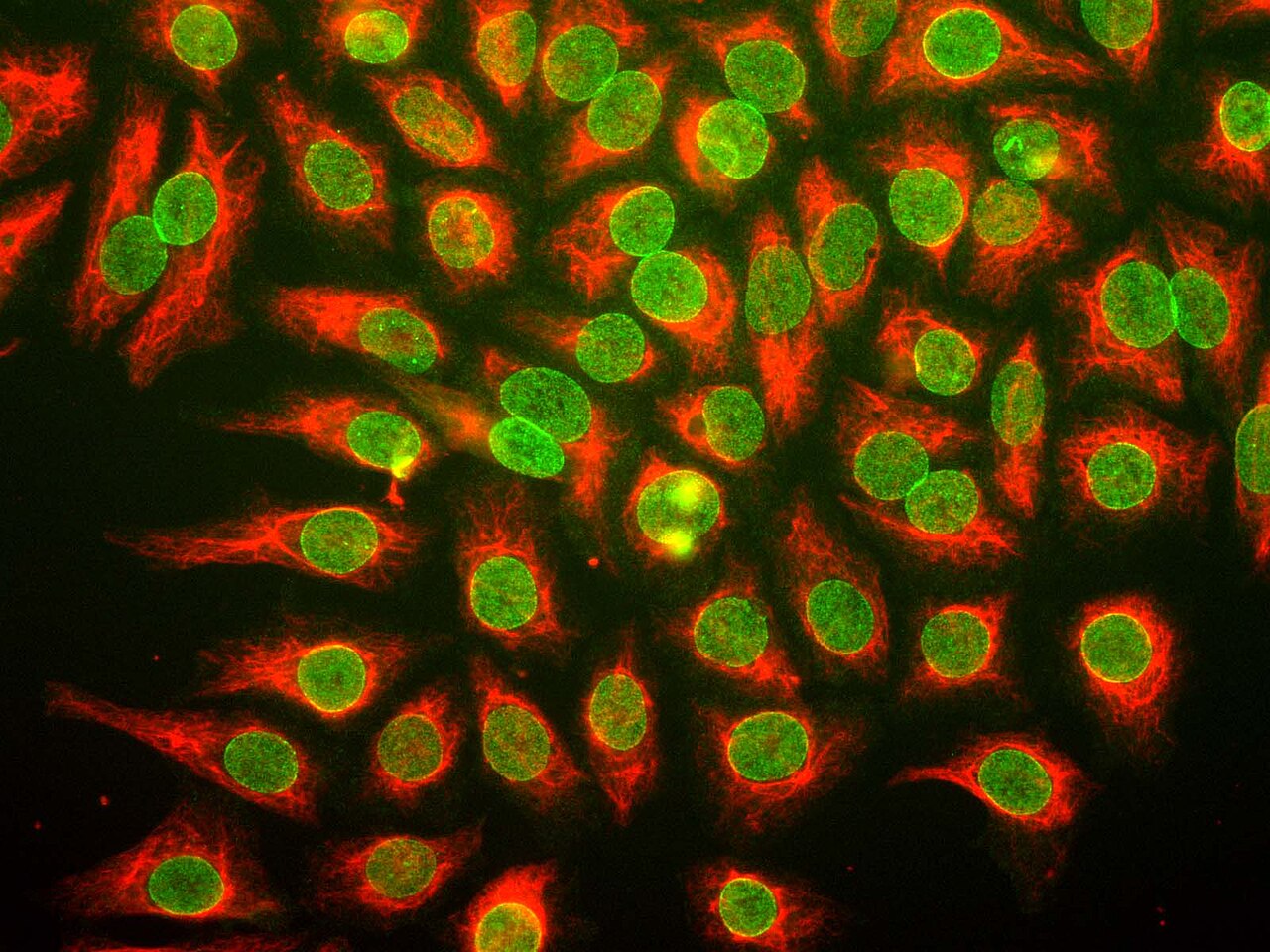
Vimentin and Nuclear Pore Complexes in HeLa Cells
GerryShaw · 27 November 2011
A 2011 fluorescence micrograph of HeLa cells stained to show internal filaments and nuclear structures, illustrating the cell line from Henrietta Lacks's tumor.
This image documents the HeLa cell line that originated from Lacks's cancer cells.
About this source
What it is
This fluorescence microscopy image was created in 2011 by GerryShaw using antibodies and imaging from EnCor Biotechnology Inc. It shows HeLa cells stained red for vimentin, a structural protein inside the cell, and green for proteins of the nuclear pore complex.
Why it matters
HeLa cells, grown from cells taken from Henrietta Lacks in 1951, have become a standard tool in laboratories studying basic cell biology, including the structures shown here. This image is a product of that long scientific use, made by researchers decades after Lacks's death, and says nothing about her life directly.
Look closer
- What two structures are highlighted in this image, and what colors represent them?
- Why might scientists choose to study these specific proteins in HeLa cells?
- What is missing from this image if you wanted to learn about Henrietta Lacks the person?
This photograph is part of the curated material that grounds the AI version of Henrietta Lacks on Humy.ai — when students ask about related events, the AI can quote it and cite it.
More Henrietta Lacks sources
Browse all 21 →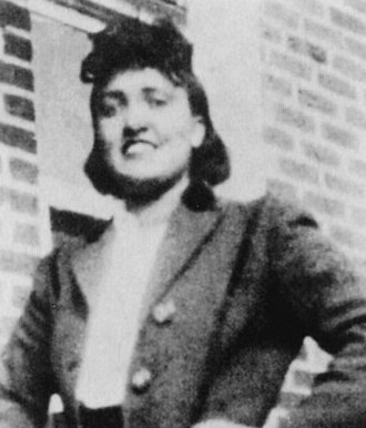 Photograph
Primary source
Portrait of Henrietta Lacks
c. 1945
 Photograph
Secondary source
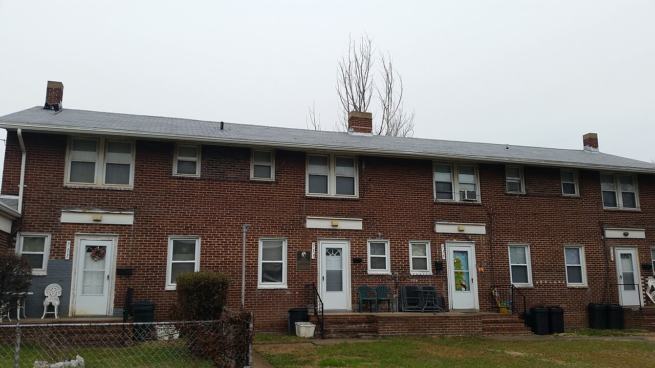
Henrietta Lacks House, Turner Station
Preservation Maryland
 Document
Primary source
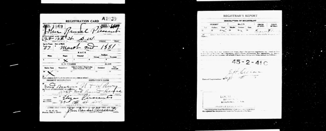
World War I Draft Registration of John Randall Pleasant
 Sculpture
Secondary source
Statue of Henrietta Lacks
Helen Wilson-Roe · 2021
Free account
Read every Henrietta Lacks source
Sign up free →
Ready when you are
Start chatting with Henrietta Lacks now.
20 messages free · No credit card · Available in 50+ languages.